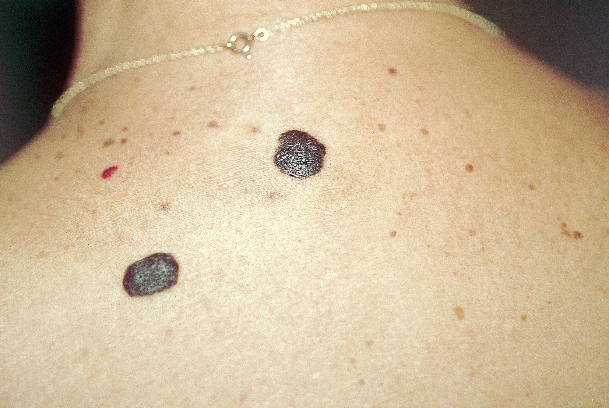
Healthremedy123.com – The skin disease Verrucous Seborrheic Keratosis is often associated with alopecia. Seborrheic keratoses are tan or black in color and can range in size from a tiny pimple to a two-centimeter (1-inch) mass. Seborrheic keratoses can be difficult to distinguish from other skin conditions, including warts and basal cell carcinoma. They are not contagious, though, and are usually accompanied by a similar appearance to warts.
Seborrheic Keratosis Consists of Sheets of Small Cells
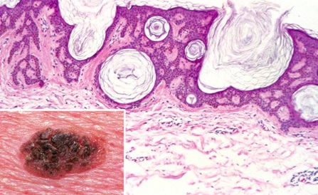
Most people experience a single or a few lesions. Those that are multiple have a condition called Leser-Trelat. The cause is unknown but has been linked to an overproduction of alpha-transforming growth factor (TGF), which binds to epidermal growth factor receptors. Seborrheic keratoses are comprised of sheets of small cells similar to basal cells.
In an 1889 study, Dr. Richard L. Baer and others described a case of a woman with seborrheic keratosis. She had long hair that covered her lesion, and she refused to be photographed with her family or friends. Interestingly, her patient had one sibling with the same type of lesion, and both had his seborrheic keratosis removed.

A 73-year-old Caucasian man presented to dermatology with a growth on his left parietal scalp that had enlarged, was bleeding, and was intermittently painful. The doctor diagnosed the growth as verrucous seborrheic keratosis and noted that the patient had no history of immunosuppression. The growth was approximately 5.2 cm x 3.8 cm.
Effects of Hydroperoxides on Seborrheic Keratosis
A series of studies conducted by Drs. Sutton, Richard L., and Szmanski, Frederick J., have investigated the role of hydrogen peroxide in seborrheic keratosis. They published a report on their studies in the Journal of Pediatric Surgery. In another study, Drs. Oliver, T. H., and Murphy, D. V., studied the effects of hydroperoxide on seborrheic keratosis and other skin diseases.
On examination, a patient presented with multiple acanthotic lesions ranging in size from 91 mm to 65 mm. Several of the lesions were ulcerated and papillomatosis. An incisional biopsy was done to rule out other conditions such as a giant SK or congenital nevus. The diagnosis was confirmed by a biopsy of the lesions, which revealed basaloid cells, epidermal hyperkeratosis, and acanthosis.

After treatment, the patient may experience a faint pink coloration on the skin, which should fade over time. In all, the patient tolerated the procedure well. The only two subjects with unusual sensitivity to the treatment sought an early application of a neutralizing composition. The most notable improvement was a complete absence of the keratosis after three years. It is not known whether the lesions will return after treatment.
Treatment for Seborrheic Keratoses
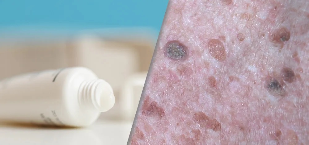
Treatment for Seborrheic Keratose includes a hydrogen peroxide solution that is at least 23 percent concentration. It also contains one or more compounds from a group of substances known as reactive oxygen species. Hydrogen peroxide, benzoyl peroxide, and alloy peroxide are particularly effective in eliminating seborrheic keratoses.
In addition to irritated SK, the condition can mimic other dermatoses. The diagnosis of SK is usually not straightforward, and complete excision and histological analysis are necessary. The patient’s presenting symptoms should be assessed to determine whether they are a true case of Verrucous Seborrheic Keratosis. A surgical removal is an option for larger lesions.

Additional treatment for Seborrheic Keratose is a topical composition that contains an ingenol compound. This compound is absorbed into the skin by the sebaceous gland and results in a reduction in the severity of symptoms. Some treatments are more effective than others, and the method described herein is an ideal treatment for Seborrheic Keratosis.
Reference:


