
Healthremedy123.com – There are two types of vessels in the thorax: the arteries and veins. The arteries and veins supply the intercostal spaces. The superior intercostal artery originates at the thoracic aorta, while the latter arises in the upper thoracic area. There are nine pairs of posterior intercostal arteries, and the last one gives branches to the ribs and skin.
Superior Intercostal Artery
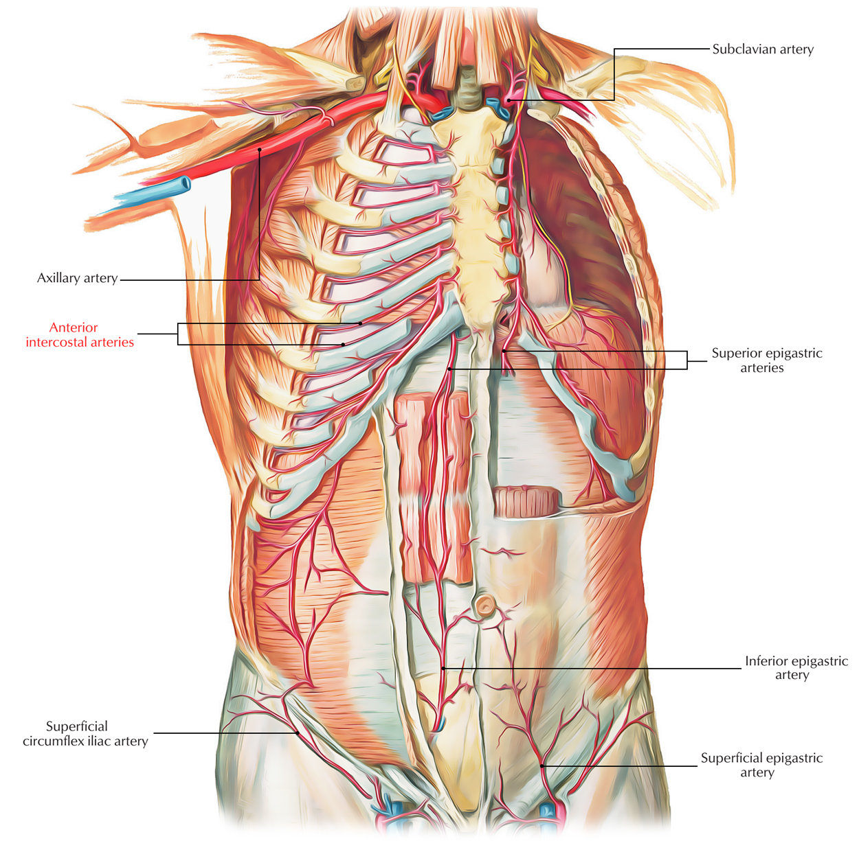
The superior intercostal artery arises from the descending branch of the internal thoracic artery. This artery enters the thorax from the medial side of the first rib, and provides blood to the second, third, and fourth intercostal spaces. The inferior intercostal artery supplies the triceps and the thymus. The aorta and veins of the axilla and sacrum supply the internal organs and the chest wall.
The lower intercostal artery comes from the descending thoracic artery and crosses the vertebral bodies and anterior longitudinal ligaments. It ends opposite the last intercostal space and supplies the 7th, eighth, and ninth intercostal spaces. The right aortic intercostal artery is longer than the left one and is situated behind the thoracic duct. It passes behind the azygos vein and is covered by the pleura of the right thoracic cavity.
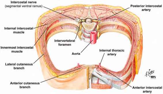
The posterior intercostal artery derives from the thoracic artery and runs behind the inferior border of each rib. The needle must pass just above the superior border of the inferior rib to ensure safe access. However, this relationship is less reliable than previously believed. Wraight et al. documented that the position of the neurovascular bundle in the superior thoracic artery varied from 50% above to 43% below the inferior border.
Posterior Intercostal Arteries Originate from the Thoracic Arteries
The posterior intercostal artery derives from the thoracic artery and the T1 nerve. It passes in front of the first rib on the right side. Its medial part lies behind the azygos artery and esophagus. Its branches are connected to the T1 nerve. Both of these vessels provide blood flow to the chest. These vessels are vital for breathing, but they also supply oxygen and nutrients to the lungs.
The aortic intercostal artery is the shortest of the intercostal arteries. It originates from the thoracic aorta and extends horizontally between the ribs. It supplies the chest wall with blood from the azygos artery. It is not a common source of coronary artery disease, but it is important for the health of the heart.

The internal thoracic artery carries blood to the intercostal arteries. The inferior intercostal artery is connected to the anterior intercostal artery. The inferior intercostal artery is found in the upper right rib. Its superior intercostal artery passes the T1 nerve and the T2 nerve. The superior intercostal arteries are the most numerous in the body. Its branches supply the most important parts of the thorax.
Internal Thoracic Artery is the Main Artery in the Chest
The internal thoracic artery is the main artery in the chest. It comes from the aorta behind the ribs. The descending branch of the artery extends behind the thoracic aorta. It continues to the sternum and the pectoralis major. It passes behind the first six intercostal cartilages. It then goes in front of the transversus thoracis muscle.
The thoracic aorta and its branches supply the intercostal arteries. The posterior intercostal artery is a branch of the aorta. The superior intercostal artery passes the front of the first rib, and the T1 nerve. The anterior and posterior aorta are connected by a branch called the azygosa. This descending thoracic artery and the right azygos are the only two arteries that supply the lower thoracic aorta.
The posterior intercostal artery supplies the right thoracic wall. It drains to the superior vena cava through the left thoracic aorta. The posterior intercostal artery is the most common artery in the body. It is the most common vein in the thoracic area. It has a median length of 3 cm, and connects to the aorta and apex of the spine.
Reference: