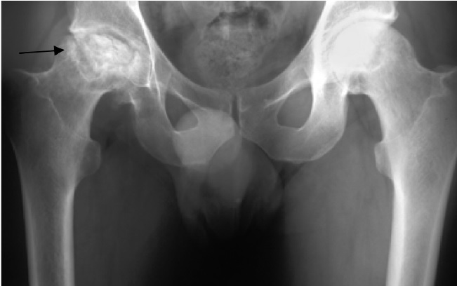
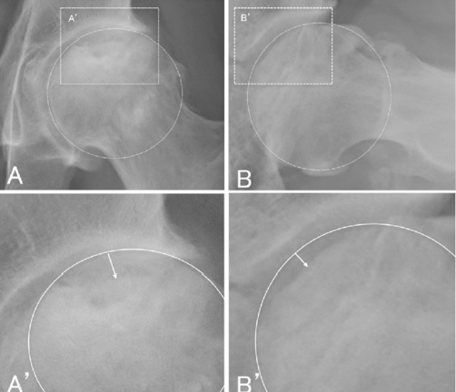
Healthremedy123.com – The Steinberg gradiHead ng system is a widely used system for femoral head avascular necrosis (ONFH). It has seven stages based on radiographic appearance and quantifies involvement of the lateral femoral head. It allows direct comparison between series, which can help diagnose and treat ONFH. The grading system is also based on the severity of disease. The different stages are indicated by the presence or absence of symptoms, subchondral fracture, and sclerosis.
Disease Progression Depends on Location and Size
The progression of the disease is dependent on its location and size. The best way to determine whether a patient has a stage I or stage II disease is to undergo an MRI. An MRI can identify the location of the lesion, which is critical in determining the prognosis. A biopsy will reveal if the disease has progressed to the next stage. If an MRI reveals the presence of a lesion, treatment options will be discussed.
The progressive stages of Avascular Necrosis of the femoral head can be seen on an MRI. The lesion may be present in any part of the head, such as the anterolateral or central part of the femur. The stage at which the disease reaches this stage is called thoracic osteonecrosis (ONFH). Avascular necrosis of the thighbone is a condition that affects one or more femur bones.

The AVNFH lesion in stage II showed an increase in size and area. In stage III, the AVNFH volume was greater than 15% of the thighbone in the group that advanced to the THA. There was no difference between the groups in the location of the necrotic lesion. In all cases, THA was necessary for patients to reach stage III. However, the vascular graft was the most commonly used.
AVNFH has Three Phases
Besides the AVNFH, there are other stages of AVNFH. The AVNFH is the first stage. The AVNFH has three phases: precollapse, early collapse, and late collapse. The final stage has no specific stage. In addition to the Oxford Bone Age stage, AVNFH is considered asymptomatic. This means that it has not reached a full maturity stage yet.
The AVNFH stage is the most severe stage of AVN. The femoral head is at the point when the vascular supply to the femur is compromised, resulting in the death of bone cells. The affected femoral head is then prone to arthritis and the patient develops pain and stiffness. It is important to seek medical treatment for AVNFH to avoid further complications and to prevent further deformity.

Despite the prevalence of this ailment, there is no clear cut diagnosis for this disorder. However, MRI scans can provide insight into early femoral head development and its relation to underlying bone age. Earlier stages of the condition can be diagnosed by examining the anterolateral femoral head. The earliest stages can be detected with the help of a fracture line. Moreover, there is a possibility of osteonecrosis without mechanical insufficiency.
X-Rays are Helpful in Determining the Degree of Avascular Necrosis
The earliest stages of ONFH are the pre-collapse, early collapse, and late collapse. During the last stages of ONFH, the patient may experience loss of range of motion, while the other stage may be in a stable condition. X-rays are helpful in determining the extent of avascular necrosis. They also show whether the femoral head is fully formed. If the latter stage is present, an MRI can help the doctor determine the stage of the ailment.
The earliest stage of ONFH occurs when the femoral head is not fully formed. The earliest stage is the first stage of the condition, followed by the second and third stages. Its progression is gradual and the patient may have a history of pain. In case of an early collapse, the patient may have a faint sclerotic band in the femoral neck. It is important to consider the patient’s pain history and underlying condition.

The stage of AVN depends on the causes of the femoral head. Most cases, the femoral head is decompressed with a core decompression. The femoral neck and the acetabular portion of the femur may be collapsed due to a traumatic event or a bacterial infection. Surgical interventions are recommended only if the AVN is small and does not cause a significant amount of pain.


